Dr. Harris’s practice is centered on hip and knee replacement, from a first-time joint replacement to more involved cases. That includes patients whose implants were placed years ago and are now wearing out, and those coming for a second opinion on a hip or knee that isn’t doing well.
Andrew B. Harris, MD · Last reviewed September 2026
Knowing what each operation involves, who it’s for, and how the options compare helps you make a confident, well-informed decision about your hip or knee.
Most hip and knee arthritis is managed first without an operation: activity modification, physical therapy, weight management, anti-inflammatory medication, and injections. Joint replacement enters the conversation when those measures no longer provide enough relief to do the things you care about: walking without planning around pain, sleeping through the night, keeping up with family.
The sections below explain each procedure clearly: what it involves, when it’s considered, and how the options differ. Every recommendation is individualized based on your symptoms, examination, and imaging. There is no one-size-fits-all operation.
Full details: anterior approach hip replacement →
Full details: STAR approach hip replacement →
The hip is a ball-and-socket joint. When the smooth cartilage lining the joint wears away, most often from osteoarthritis, the common “wear-and-tear” form of arthritis, bone begins to rub on bone, causing pain, stiffness, and a shrinking radius of activity. A hip replacement removes the worn ball and socket and replaces them with implants sized and positioned to recreate your specific anatomy.
Hip replacement has been called “the operation of the century”: for most patients it means substantial pain relief and a return to walking, cycling, golf, travel, and daily life without thinking about the hip.
Dr. Harris plans the operation from your own imaging and routinely uses robotic assistance or computer navigation to help position the implants, fitting the socket to your anatomy.
There are two main surgical approaches to the hip. Both are well-established approaches with similar results in most studies, and Dr. Harris performs both. Which one suits a given patient depends on anatomy, imaging, and health history rather than a single fixed philosophy.
From the front of the hip · Dr. Harris’s preferred approach
This is the approach Dr. Harris prefers for most hip replacements. It reaches the hip by working between the muscles rather than cutting through them, which for many patients can mean less discomfort early on and a quicker return to walking. Some studies have found lower dislocation rates with the anterior approach than with a traditional posterior approach, while others have found them similar. When your anatomy is a good fit, he can perform it through a “bikini” incision, a shorter cut that follows the natural crease of the skin, leaving a lower, more discreet scar.
A modern, tissue-sparing approach from the back of the hip
Dr. Harris performs the STAR approach (Superior Transverse Anatomic Reconstruction), an updated version of the traditional approach from the back of the hip. It gives a clear, complete view of the joint while preserving the muscles and tendons that help keep the hip steady, structures that older techniques often divided.
Full details: partial knee replacement →Full details: patellofemoral joint replacement →Full details: robotic-assisted knee replacement →
Despite the name, a knee replacement is closer to a resurfacing. The worn cartilage and a thin layer of bone are removed from the ends of the thigh bone and shin bone, then capped with smooth metal and plastic surfaces, much like a dentist crowns a damaged tooth. Your own muscles and most ligaments remain, which is why rehabilitation is so central to a good result.
It’s considered when arthritis pain limits daily life despite non-surgical treatment, most often from osteoarthritis (the everyday “wear-and-tear” arthritis), but also inflammatory arthritis or arthritis that develops years after a knee injury.
Most knee replacements are done through a standard approach, but for some patients another option is a subvastus (muscle-sparing) technique that slips beneath the quadriceps muscle instead of cutting through the tendon. Whether it fits depends on your anatomy, and Dr. Harris will discuss it when it’s appropriate.
Most patients are up and walking the day of surgery, though recovery varies from person to person. Dr. Harris uses current multimodal techniques to manage pain and swelling afterward, keeping early recovery as comfortable as possible. Physical therapy is also an important part of the recovery process for most patients, with most of the improvement coming in the first three months.
For many knees, Dr. Harris uses robotic assistance and computer navigation to help fine-tune implant position and soft-tissue balance. Modern implants are very durable, with large registry studies showing most knee replacements still in place 25 years after surgery, and many lasting 30 years or more.
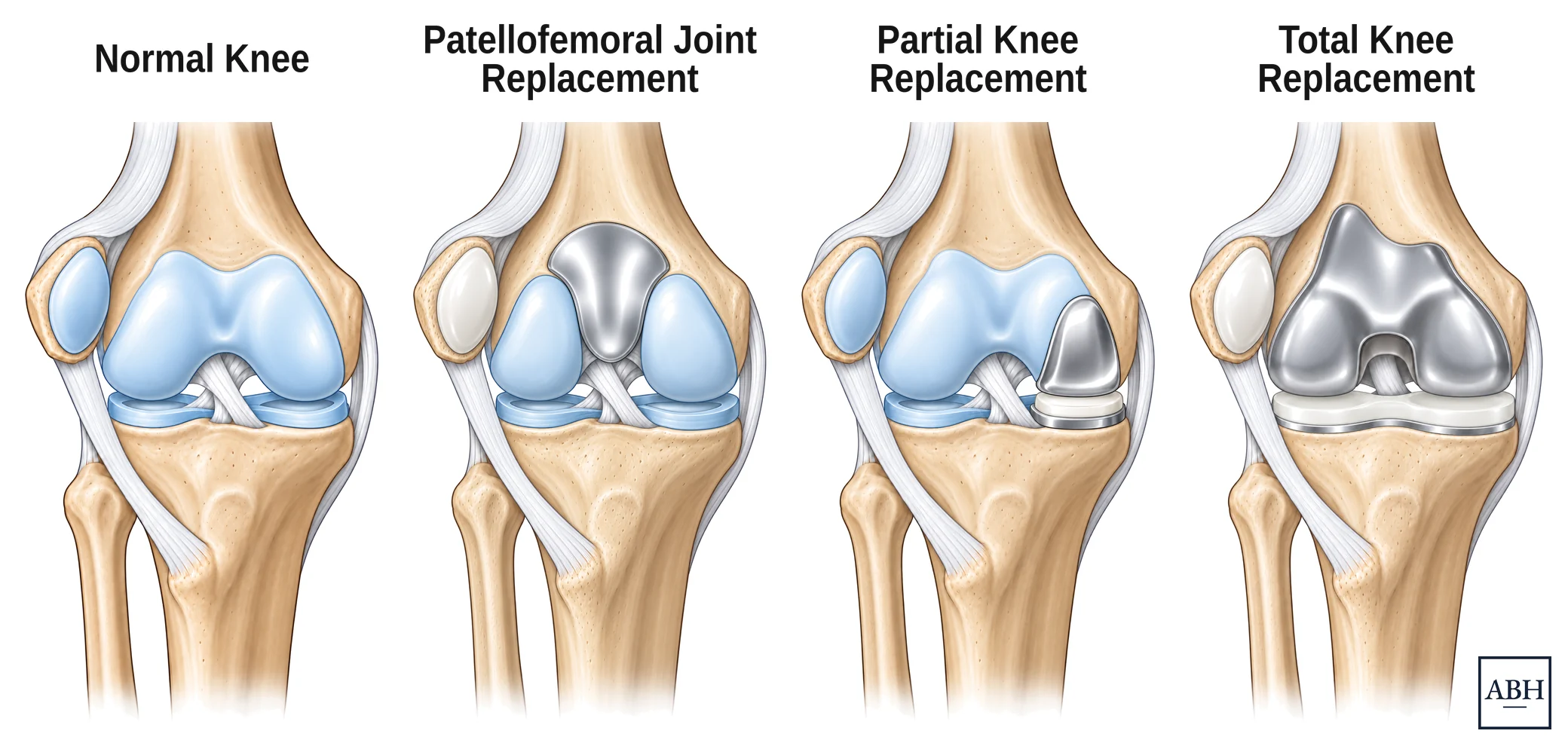
One knee, then the three operations. How much of the joint is resurfaced follows how much of it is worn.
For arthritis throughout the knee
All three compartments of the knee are resurfaced. This is the most common knee replacement and the right choice when wear involves most of the joint. It offers lasting pain relief for most patients, though some have ongoing pain or stiffness.
For wear limited to one part of the knee
Only the damaged part of the knee is resurfaced; healthy cartilage and all the ligaments are preserved. Depending on where the wear is, this may be a medial partial knee replacement (inner side), a lateral partial knee replacement (outer side), or a patellofemoral joint replacement (behind the kneecap). In carefully selected knees, a partial knee replacement can feel more natural and recover faster, but is more likely than a total knee replacement to need further surgery later. More on partial knee replacement.
Revision joint replacement surgery is some of the most technically demanding work in hip and knee surgery, and Dr. Harris’s fellowship training included complex revision surgery.
Revision surgery removes some or all of an existing implant and rebuilds the joint, often with specialized components designed for bone that has changed since the first operation. Part of Dr. Harris’s research looks at why implants fail.
The first task is understanding exactly why a joint replacement is failing, which is as important as the operation itself and shapes everything that follows. With that diagnosis in hand, Dr. Harris evaluates and treats hip and knee replacements that have failed, including implants placed by other surgeons.
Any of these can be a reason to have a hip or knee replacement evaluated:
Full details: robotic-assisted hip replacement →
Full details: robotic-assisted knee replacement →
Dr. Harris routinely uses robotic assistance or computer navigation in his hip and knee replacements. Each robotic-assisted operation is planned in advance from the patient’s own imaging, and the system helps carry that plan out precisely in the operating room. He has trained on four of the systems in current use for the knee: Mako, ROSA, CORI and VELYS. For hip replacement he uses the Mako system.
The technology is a tool rather than a different operation. Patients can do very well with a joint replacement done with robotic assistance and with one done without it. What the evidence shows consistently is more accurate implant position, without a proven difference in long-term outcomes.
On the hip side, robotic assistance and navigation are used with both the direct anterior approach and the STAR approach.
Mako is a trademark of Stryker; ROSA of Zimmer Biomet; CORI of Smith+Nephew; and VELYS of DePuy Synthes. Naming these systems does not imply endorsement by, or affiliation with, their makers. Payments from device makers to physicians, including meals, are reported publicly through CMS Open Payments.
General information for patients and clinicians, not a substitute for advice from your own surgeon.
If you are weighing whether surgery makes sense, or want a second opinion, you are welcome to get in touch.